Lasers, Magnetic Stimulation and a Robotic Arm: How Researchers at HSE University Study the Brain

The Institute for Cognitive Neuroscience (ICN) at HSE University has recently added state-of-the-art laboratory equipment to its range of tools for studying brain function. The News Service visited the Institute to learn more about the uses of infrared lasers, optical tomography and a unique robotic arm, as well as why research into vascular tone is important, which parts of the brain can be stimulated to make people more generous, and how the Institute’s research can help treat diseases.
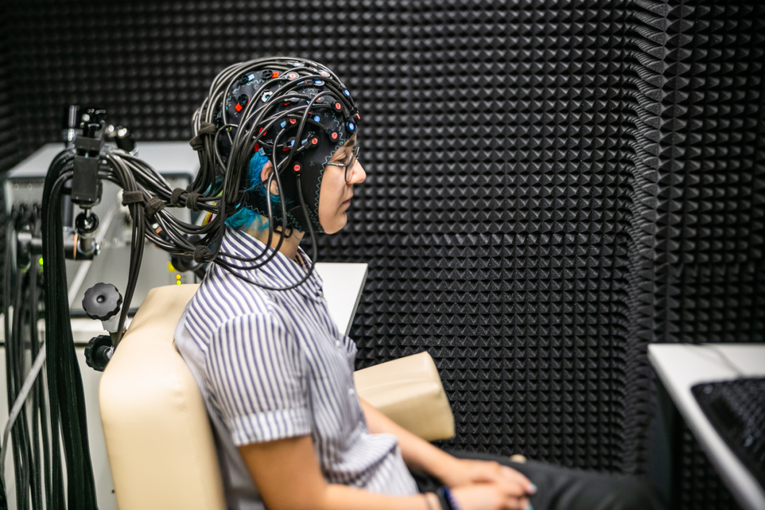
The Functional Optical Tomography laboratory looks like a medical room for EEG examinations. A comfortable chair stands next to a computer, which is connected to a ‘helmet’ made up of wires and dozens of sensors that envelop the test subject’s head. The room containing the optical tomograph itself is shielded by a ‘shell’ of special material that protects the device from external interference.
Aleksei Gorin, Junior Research Fellow at the Centre for Cognition and Decision Making at the ICN, told the HSE News Service how the process works. Special sensors conduct optical pulses from infrared lasers to detect differences in vascular tone in the veins and arteries. This in turn is used to measure oxygen absorption levels in various parts of the brain associated with logical problem solving and mental, sensory and motor activity. This allows researchers to study the brain's capabilities and its ability to respond to different tasks with more precision.
When our News Service reporter tested the equipment by reading a poem to himself, there was a drop in signal strength from the sensors monitoring the concentration of oxygenated blood. This, explained laboratory staff, reflects an increase in oxygen consumption in the brain.
The new equipment can also be used to study how the brain works when making certain decisions
The technology makes it possible to investigate certain kinds of brain activity and their impact on the brain's computational (and other) abilities. Optical tomography research also has medical applications, namely for the early diagnosis of heart and brain diseases.
In the absence of a perfect tool for studying the brain, a variety of experimental methods are required to get a clearer picture, Mr. Gorin explained. Researchers can conduct detailed behavioural research on the influence of various brain processes on human behaviour, and predict with a reasonable degree of accuracy which decisions an individual might make while affected by one process or another. ‘You can also give the brain a task and see how external factors affect how the brain works when performing decision-making tasks, sensory tasks or attention tasks. You can also learn more about how the brain works when someone is resting and relaxing’, he said.
Mr. Gorin also said that the Laboratory is interested in conducting interdisciplinary research with economists and medical professionals. Over the last two years, the Laboratory’s staff has expanded to include other specialists, including neurologists and psychiatrists. This has helped broaden the scope of its brain research.
Tatiana Chernyakova, Research Assistant at the International Laboratory of Social Neurobiology, said that the Laboratory’s plans for the near future include neuromarketing research and two cognitive experiments with the new equipment, using a combination of MRI and optical tomography to obtain new data.
The Transcranial Magnetic Stimulation (TMS) laboratory has been overseeing the operation of a state-of-the-art stimulation device capable of activating parts of the brain. The device is installed next to a chair similar to one found in a dentist's office. A unique robotic arm (manufactured in France as there is currently no Russian equivalent) is placed above the head of a test subject and adapts to their movements. The accuracy of the robotic arm can be checked using a computer containing information about the area to be stimulated.
Matteo Feurra, Leading Research Fellow at the ICN Centre for Cognition and Decision Making, explains that if the subject makes any sudden movements for any reason (such as feeling unwell), the robotic arm deactivates the pulses and withdraws to a safe distance.
During the stimulation process, the robotic arm moves closer to the subject’s head, on which a reference bar has been placed to keep track of its position. The robot arm moves to match even the slightest movements of the head and sends an impulse to the target area of the brain. This induces the desired reaction, such as the contraction of muscles in the arm or fingers. While this result is visible in the test subject, the induced potential is also recorded and displayed by the computer.
Oksana Zinchenko, Research Fellow at the International Laboratory for Social Neurobiology, noted that stimulation has both research and medical applications. Once such use is the restoration of cells located around lesions or in the opposite hemisphere to an area affected by a stroke. Research Fellow Ainur Ragimova explained that transcranial magnetic stimulation can be used to treat depression and cervical dystonia, a disease that causes neck spasms, difficulty turning the neck and pain.
The TMS laboratory has a dedicated room for conducting complex research into brain stimulation and recording its effects. It is also shielded by a 'shell' for maximum signal clarity and isolation from external influences.
Areas of interest for this research include decision-making, social decisions, the speech areas of the brain, and memory. Ms. Zinchenko also explained that TMS is used to study the prefrontal cortex of the frontal lobe, which is responsible for selfish or prosocial motivations when deciding how to allocate resources—such as choosing whether or not to share points in a social game.
Using TMS to block the prefrontal cortex makes a person's behaviour more prosocial—they become more generous
Shutting down a group of neurons makes it possible to identify certain causal relationships. Researchers can see whether shutting down or stimulating individual areas disrupts or improves performance at a given task, and thus determine which areas of the brain are responsible for said tasks. These findings can then be used to build cognitive maps.
The laboratories and various research methods are available to specialists from different departments of the Institute for Cognitive Neurosciences, including foreign scientists such as Iiro Jääskeläinen, Academic Advisor of the International Laboratory of Social Neurobiology, Maria Del Carmen Herrojo-Ruiz, Leading Research Fellow at the ICN, Vladimir Djurdjevic, Research Fellow of the Centre for Cognition and Decision Making, Matteo Feurra and others.
Aleksei Gorin
Vladimir Djurdjevic
Ainur Ragimova
Tatiana Chernyakova
See also:
'We Are Creating the Medicine of the Future'
Dr Gerwin Schalk is a professor at Fudan University in Shanghai and a partner of the HSE Centre for Language and Brain within the framework of the strategic project 'Human Brain Resilience.' Dr Schalk is known as the creator of BCI2000, a non-commercial general-purpose brain-computer interface system. In this interview, he discusses modern neural interfaces, methods for post-stroke rehabilitation, a novel approach to neurosurgery, and shares his vision for the future of neurotechnology.
Smoking Habit Affects Response to False Feedback
A team of scientists at HSE University, in collaboration with the Institute of Higher Nervous Activity and Neurophysiology of the Russian Academy of Sciences, studied how people respond to deception when under stress and cognitive load. The study revealed that smoking habits interfere with performance on cognitive tasks involving memory and attention and impairs a person’s ability to detect deception. The study findings have been published in Frontiers in Neuroscience.
'Neurotechnologies Are Already Helping Individuals with Language Disorders'
On November 4-6, as part of Inventing the Future International Symposium hosted by the National Centre RUSSIA, the HSE Centre for Language and Brain facilitated a discussion titled 'Evolution of the Brain: How Does the World Change Us?' Researchers from the country's leading universities, along with health professionals and neuroscience popularisers, discussed specific aspects of human brain function.
‘Scientists Work to Make This World a Better Place’
Federico Gallo is a Research Fellow at the Centre for Cognition and Decision Making of the HSE Institute for Cognitive Research. In 2023, he won the Award for Special Achievements in Career and Public Life Among Foreign Alumni of HSE University. In this interview, Federico discusses how he entered science and why he chose to stay, and shares a secret to effective protection against cognitive decline in old age.
'Science Is Akin to Creativity, as It Requires Constantly Generating Ideas'
Olga Buivolova investigates post-stroke language impairments and aims to ensure that scientific breakthroughs reach those who need them. In this interview with the HSE Young Scientists project, she spoke about the unique Russian Aphasia Test and helping people with aphasia, and about her place of power in Skhodnensky district.
Neuroscientists from HSE University Learn to Predict Human Behaviour by Their Facial Expressions
Researchers at the Institute for Cognitive Neuroscience at HSE University are using automatic emotion recognition technologies to study charitable behaviour. In an experiment, scientists presented 45 participants with photographs of dogs in need and invited them to make donations to support these animals. Emotional reactions to the images were determined through facial activity using the FaceReader program. It turned out that the stronger the participants felt sadness and anger, the more money they were willing to donate to charity funds, regardless of their personal financial well-being. The study was published in the journal Heliyon.
Spelling Sensitivity in Russian Speakers Develops by Early Adolescence
Scientists at the RAS Institute of Higher Nervous Activity and Neurophysiology and HSE University have uncovered how the foundations of literacy develop in the brain. To achieve this, they compared error recognition processes across three age groups: children aged 8 to 10, early adolescents aged 11 to 14, and adults. The experiment revealed that a child's sensitivity to spelling errors first emerges in primary school and continues to develop well into the teenage years, at least until age 14. Before that age, children are less adept at recognising misspelled words compared to older teenagers and adults. The study findings have beenpublished in Scientific Reports .
Meditation Can Cause Increased Tension in the Body
Researchers at the HSE Centre for Bioelectric Interfaces have studied how physiological parameters change in individuals who start practicing meditation. It turns out that when novices learn meditation, they do not experience relaxation but tend towards increased physical tension instead. This may be the reason why many beginners give up on practicing meditation. The study findings have been published in Scientific Reports.
Processing Temporal Information Requires Brain Activation
HSE scientists used magnetoencephalography and magnetic resonance imaging to study how people store and process temporal and spatial information in their working memory. The experiment has demonstrated that dealing with temporal information is more challenging for the brain than handling spatial information. The brain expends more resources when processing temporal data and needs to employ additional coding using 'spatial' cues. The paper has been published in the Journal of Cognitive Neuroscience.
Neuroscientists Inflict 'Damage' on Computational Model of Human Brain
An international team of researchers, including neuroscientists at HSE University, has developed a computational model for simulating semantic dementia, a severe neurodegenerative condition that progressively deprives patients of their ability to comprehend the meaning of words. The neural network model represents processes occurring in the brain regions critical for language function. The results indicate that initially, the patient's brain forgets the meanings of object-related words, followed by action-related words. Additionally, the degradation of white matter tends to produce more severe language impairments than the decay of grey matter. The study findings have been published in Scientific Reports.


